
The lymph nodes are part of the lymphatic system. The lymphatic system helps fight infections and is made up of lymph vessels, lymph fluid, lymph nodes, bone marrow and lymphatic organs (thymus, adenoid, tonsil and spleen).
Lymph vessels are thin tubes, similar to blood vessels. They collect and move lymph fluid away from tissues into the lymph nodes and back to the heart. Lymph nodes are small bean-shaped organs of lymphatic tissue. Cancer cells may migrate through lymphatic channels to other parts of the body.
Lymph fluid from the breast, skin of the upper limbs and other nearby tissues drains into the axillary lymph nodes. Lymph can carry cancer cells from these areas into the axillary lymph nodes. In the early stages, you cannot usually feel the cancer in the axillary lymph nodes. In more advanced stages of cancer, you may feel a lump in the armpit as the axillary lymph nodes get bigger.
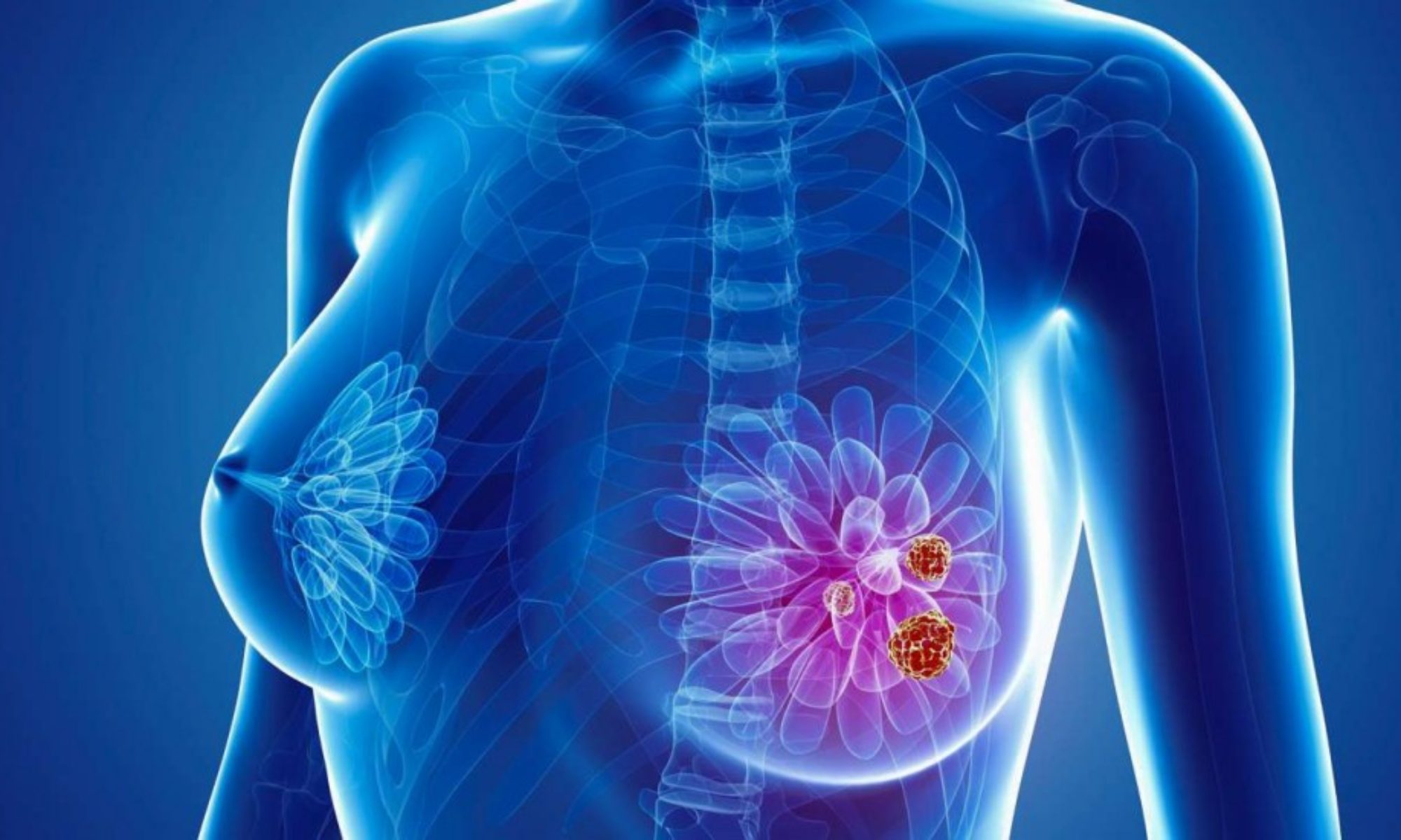
The most common type of cancer that spreads to the axillary lymph nodes is breast cancer. Other cancers may also spread to the axillary lymph nodes, e.g. melanoma.
Sentinel Lymph Node Biopsy (SLNB)
Previously, all patients diagnosed with breast cancer were treated with surgery to the breast and axillary node clearance (ALND; see below).
In patients with early breast cancer, where there is no obvious involvement of lymph nodes at diagnosis, patients will be offered sentinel lymph node biopsy (SLNB). This is usually performed at the same time as breast surgery. This procedure aims to identify and sample the nodes that receive the first lymphatic drainage from the breast. This is the most sensitive staging technique we have. By sampling these nodes we can determine the risk of the cancer spreading to other parts of the body, and may guide other therapies. This may include recommendations for systemic therapies (hormone and chemotherapy), radiation therapy and the need for further surgery.
How is a sentinel node biopsy done?
A radioactive substance is injected into the breast prior to surgery. During the surgery the surgeon may also inject a blue dye to assist with the visual identification of these nodes. The surgeon may then use a probe during the operation to detect these lymph nodes where the dyes accumulate. This procedure may be performed at the same time as surgery on the breast, often through a small incision under the armpit. The nodes identified using the dyes are then removed and sent to the laboratory where they are reviewed by a pathologist.
Benefits of Sentinel Lymph Node Biopsy (SLNB)?
SLNB enables doctors to accurately stage cancers and estimate risk of the tumour spreading to other parts of the body. SLNB may guide further therapies or treatment, and help predict the risk of cancer recurrence. For some patients SLNB may be therapeutic and remove tumour cells, reducing the risk of local recurrence, and may help avoid the need for more extensive surgery or treatments.
Side effects of SLNB
- Seroma is the accumulation of fluid in the arm pit at the site of surgical resection. This is caused by the disruption of lymphatic flow due to surgery. If this occurs you may notice a firm, tender swelling under the arm. This may resolve spontaneously over weeks, but if it causes symptoms then your doctor may decide to drain it with a needle. This may need to be repeated until the lymphatic fluid finds alternative drainage pathways.
- Numbness, tingling or pain at the site of surgery. This may result from injury to nerves at the surgical incision site. This usually improves with time but for some patients may be permanently altered.
- Lymphoedema or swelling of the upper limb is a risk during this procedure as it results in the disruption of lymphatic drainage. However, compared to the risk of lymphoma following ALND (see below), the risk is much lower (<5%).
- Shoulder stiffness and reduced range of movement. Patients are encouraged to move the shoulder following surgery. They may receive information from physiotherapy and breast care nurses to assist with gentle exercises to prevent and treat this condition.
- Lymphangiosarcoma is a rare cancer of the lymphatic system. It usually results from inflammation associated with chronic lymphoedema.
Axillary Lymph node Dissection (ALND)
An axillary lymph node dissection (ALND) is surgery to remove lymph nodes from the armpit (underarm or axilla). An ALND is also called axillary dissection, axillary node dissection or axillary lymphadenectomy.
Why an axillary lymph node dissection is done
An axillary lymph node dissection is done to:
- Remove lymph nodes that contain cancer
- Find out how many lymph nodes contain cancer and how much cancer has spread to them
- Reduce the chance that the cancer will come back (recur)
- Help doctors plan further treatment
How an axillary lymph node dissection is done
An ALND is done under general anesthetic in a hospital operating room. It may be done at the same time as surgery for the primary cancer (such as during surgery to treat breast cancer).
The surgeon makes a cut (incision) under the arm and removes 10–40 lymph nodes from level I and level II. Level III lymph nodes are not usually removed because this does not improve survival and increases the chances of side effects. However, level III lymph nodes may be removed if the cancer has spread to the lymph nodes which may be detected on imaging tests (such as an ultrasound, a CT scan or an MRI) or if they are suspicious at time of surgery.
The lymph nodes and any other tissue removed during surgery are sent to a lab to be examined by a pathologist.
After removing the lymph nodes, the surgeon places a small tube (drain) and closes the cut with stitches or staples. A drainage bag is attached to the end of the tube to collect fluid draining from the area. This reduces the chance of fluid building up and improves healing. The drain is left in place until there is little drainage (usually 5-10 days). Community nurses are often involved to help with the management of surgical drains.
People who have an ALND are usually sent home 1–2 days after surgery. You may be given:
- Pain-relieving medicine
- Instructions on caring for and dressing the wound
- Information about how to manage the drainage bag and tube
- Advice on how much and which types of activity you can do after surgery
- A follow-up appointment to see the surgeon in 1–2 weeks
- Information about which symptoms and side effects you should report
Side effects
Side effects can happen any time during, immediately after or a few days or weeks after an ALND. Sometimes late side effects develop after months or years. Most side effects go away on their own or can be treated, but some may last a long-time or become permanent.
Tell the healthcare team if you have these side effects or others you think may be from an ALND:
- Signs of infection, such as pain, redness, pus, discharge or fever
- A collection of fluid under the skin (seroma) in the armpit near the cut
- A swollen and tight-feeling arm
- Stiffness or trouble moving the arm or shoulder
- Changes in sensation, such as pain or numbness
- Chronic pain (may be caused by damage to the nerves in the armpit)
- Axillary web syndrome (AWS, also called lymphatic cording), which is when cords of scar tissue develop in the lymph vessels from the armpit to the elbow and causes pain, tightness in the arm and trouble moving the shoulder
The healthcare team may give you antibiotics to prevent or treat an infection, or they may drain a buildup of fluid.
Swelling may be due to a buildup of lymph fluid in the soft tissues (called lymphedema). Lymphedema can happen any time after lymph nodes are removed, but it is more common with an ALND. The chance of developing lymphedema increases with the number of lymph nodes removed and if radiation is given after an ALND. About 1 in 5 people get mild lymphedema after an ALND. A small percentage of people may get severe lymphedema. Lymphedema treatment may include massage therapy, compression garments and exercises.
What the results mean
Each lymph node removed is examined to see if it contains cancer.
- A negative lymph node has no cancer cells.
- A positive lymph node has cancer cells.
The pathologist’s report includes the type of cancer, the number of lymph nodes removed and the number of lymph nodes that have cancer cells. The report may also say if the cancer has grown through the outer covering of the lymph node (the capsule).
Doctors use the number of positive lymph nodes to help stage the cancer. They use the stage along with other information about the type and grade of the cancer to make treatment decisions and give a prognosis.
Information above adapted from: https://www.cancer.ca